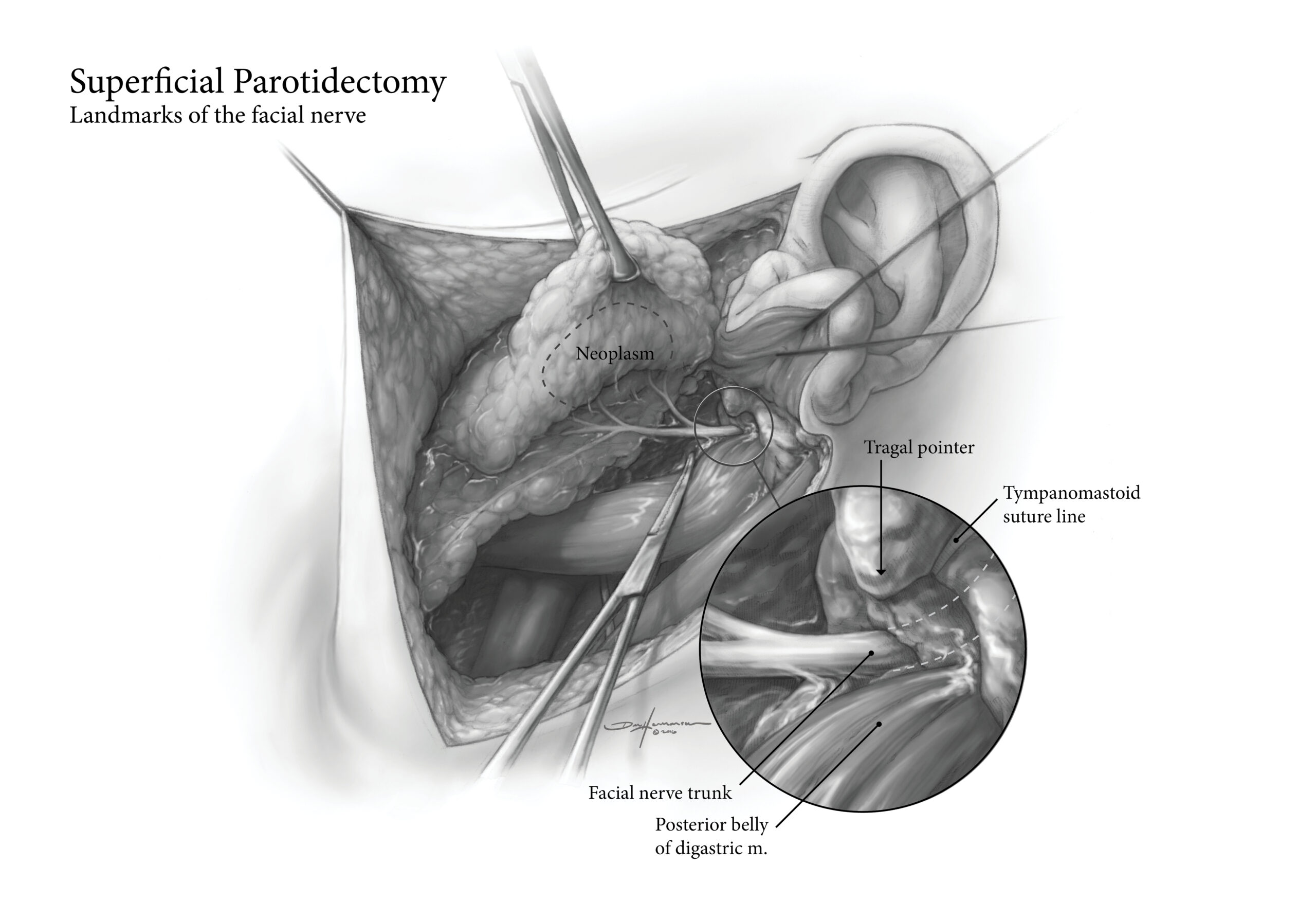
This black-and-white pencil and digital illustration provides a highly detailed anatomical depiction of a superficial parotidectomy, with a specific focus on the landmarks of the facial nerve. Designed as an educational surgical reference, the artwork combines the precision of hand-drawn textures with the clarity of digital refinement, ensuring a balance between artistic expression and medical accuracy.
This black-and-white pencil and digital illustration provides a highly detailed anatomical depiction of a superficial parotidectomy, with a specific focus on the landmarks of the facial nerve. Designed as an educational surgical reference, the artwork combines the precision of hand-drawn textures with the clarity of digital refinement, ensuring a balance between artistic expression and medical accuracy.
The composition highlights:
- Surgical exposure of the parotid gland, with the overlying tissue carefully sectioned to reveal the underlying structures.
- Key anatomical landmarks of the facial nerve, including its emergence from the stylomastoid foramen, branching within the parotid gland, and the relationship to the tragal pointer, retromandibular vein, and the posterior belly of the digastric muscle.
- Stepwise dissection techniques, with emphasis on preserving nerve integrity while excising the superficial lobe of the gland.
Shading and line weight variations are meticulously used to differentiate depth, tissue planes, and vascular structures, creating a high-contrast, instructive visualization suitable for surgical training, medical education, and reference materials. The hybrid pencil-digital approach enhances fine details, ensuring clarity in delicate areas such as the parotid fascia and nerve branches.
This illustration serves as an essential visual guide for surgeons, medical students, and educators, offering a concise yet comprehensive depiction of the procedure and its critical neurovascular landmarks.
| Color | White |
|---|


